A Year of COVID-19

Paul Whittaker • 15 March 2021

From SARS-CoV-2 to COVID-19 Pathogenesis, Vaccines and Therapeutics

On 11th March 2020, the World Health Organisation declared pandemic  status for  COVID-19, an infectious disease caused by the new coronavirus SARS-CoV-2. This virus was first isolated from airway epithelial cells of patients presenting with pneumonia of unknown cause in Wuhan, China, in December 2019. Since then, there has been a concerted global effort to characterise the virus and understand the pathophysiology of COVID-19, develop vaccines and search for drug therapies. As of 10th March 2021, 111,300 COVID-19 or SARS- CoV-2 citations can be found in  PubMed. That number is growing daily.

A year on, the aim of this article is to give an overview of what has been learned about the SARS-CoV-2 virus (Fig. 1.) and the symptomatology and pathogenesis of COVID-19 based on a review of published literature, particularly how it relates to disease severity. Specific details may change as future research expands and refines current knowledge, but hopefully this article will remain substantially accurate for the most part.

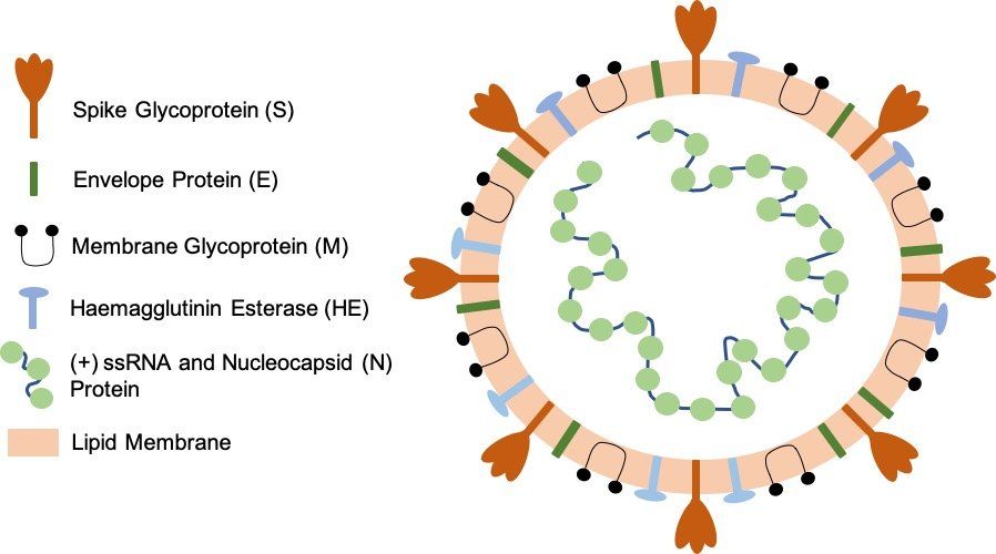
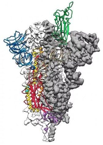
Fig. 1. SARS-CoV-2 virion particle. The coronavirus virion consists of the following structural proteins: spike glycoprotein (S), envelope protein (E), membrane glycoprotein (M), nucleocapsid protein (N) and haemagglutinin-esterase (HE). The positive-sense, single-stranded RNA genome (+ssRNA) is encapsidated by N, whereas M and E ensure its incorporation into the viral particle during the assembly process. S trimers protrude from the host-derived viral envelope and provide specificity for the cellular entry receptors ACE2 and TMPRSS2 (Fig. 2.).

TRANSMISSION AND INFECTION

Transmission and Detection

SARS-CoV-2 is spread predominantly through aerosols and airborne droplets produced by infected individuals coughing and sneezing. COVID-19 symptoms appear after an incubation period of 5 to 6 days, with peak symptoms occurring 2 to 5 days later. Virus can be detected during the incubation period by analysis of nasal or throat swab samples collected from infected individuals. A wide range of detection methods have now been developed to detect the presence of virus, with RT-PCR being the most widely used. Shedding of virus particles by infected individuals, particularly superspreaders, before symptom onset, contributes to the difficulty of controlling virus spread by public health interventions.

Infection

Infection leading to COVID-19 occurs when SARS-CoV-2 spike protein attaches to ACE2 (Fig. 2.), a cell surface protease expressed at variable levels on the surface of epithelial cells in the mouth, nose, and upper and lower airways of the lungs. After attachment to ACE2, the transmembrane protease TMPRSS2 cleaves and primes the bound spike protein to facilitate cell entry by endocytosis. Once inside the cell, the virus hijacks the host cellular machinery to replicate itself and assemble more virions. Virus particles are unpackaged and genes encoded by the viral RNA genome are translated into viral proteins by host ribosomes in the endoplasmic reticulum. Viral progeny are then assembled from the synthesised structural proteins and replicated viral RNA genome in the Golgi complex before being released from the host cell by exocytosis. One infected host cell can create up to a hundred new virions, accelerating the spread of the virus via infection of other cells in the respiratory tract.

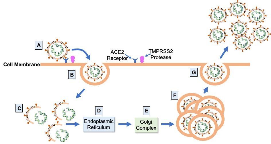
Fig. 2. SARS-CoV-2 infection cycle. Virions bind to ACE2 on the apical surface of epithelial cells in the respiratory tract via the virus S glycoprotein (A). Cleavage and priming of the bound S protein by TMPRSS2 enables endocytosis of virion particles (B). After cell entry, uncoating of the virions releases the viral genomic RNA (C) which is then translated on the ribosomes of the endoplasmic reticulum (D) to form the proteins of the viral replication and transcription complex, as well as the viral structural proteins. Translated structural proteins transit through the Golgi complex (E), where interaction with N-encapsidated, newly produced genomic RNA results in the formation of mature virions (F) ready for secretion from infected cells by exocytosis (G).

COVID-19 and the Respiratory Tract

The respiratory tract is the first line of defence against inhaled virus particles. SARS-CoV-2 shows a gradient of infectivity from the upper to the lower airways, matching the distribution of ACE2 expression levels. Cytokines released by infected airway epithelial cells and resident, plus incoming, immune cells mediate the cell-cell communication required for a tightly regulated immune response. In COVID-19, the host immune response is a critical component of disease progression and severity. Dysregulation of this response is a key feature in individuals with severe COVID-19.

COVID-19 SYMPTOMATOLOGY AND SEVERITY

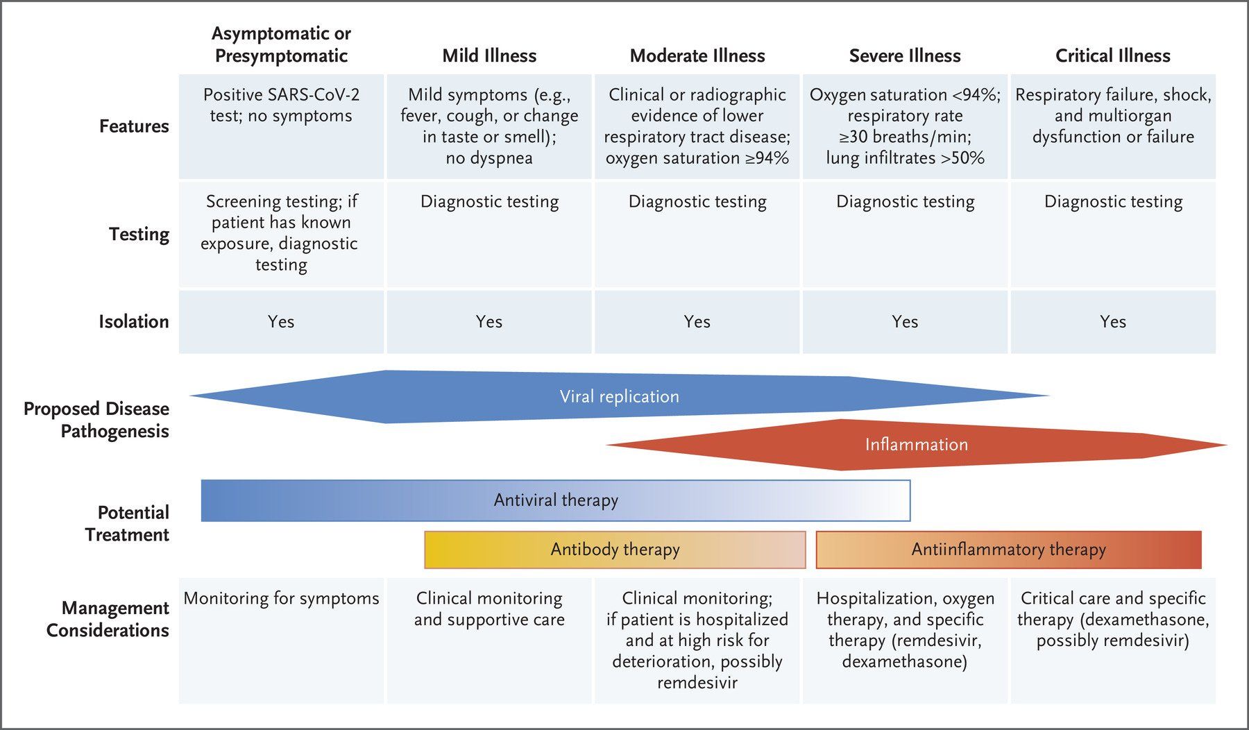
The symptoms (Fig. 3.) displayed by people infected with SARS-CoV-2 vary in type, number and severity, as well as duration (Fig. 4.). These range from asymptomatic carriage, to mild/moderate disease, to severe disease, characterised by atypical pneumonia, respiratory failure and acute respiratory distress syndrome (ARDS). The most severely affected patients tend to be older men, people from ethnic minorities and people with co-morbidities, such as obesitydiabetes and hypertension. In addition, factors such as geneticsviral load, the level of immune response and degree of lung damage will also play a part, with a predominance of different factors in different patients causing different individual clinical outcomes.

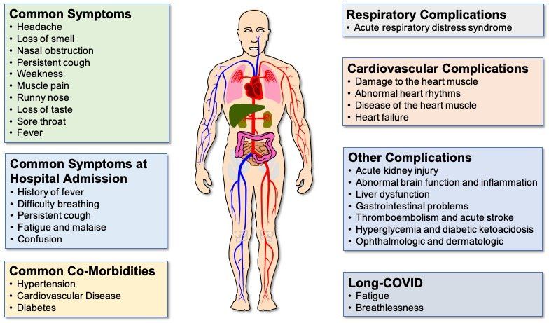
Fig. 3. COVID-19 symptoms and complications. Although fever, persistent cough and loss of taste and/or smell are the most common COVID-19 symptoms reported, some patients display a number of different symptoms. Difficulty breathing is a predominant feature of the majority of COVID-19 patients who are hospitalised. Common co-morbidities are heart disease, high blood pressure and diabetes. Although the lung is the primary site of COVID-19 pathology, complications affecting a range of extra-pulmonary organs are often seen in patients with severe disease, particularly those with co-morbidities causing endothelial cell dysfunction. Even in patients with mild/moderate disease, some patients experience long COVID, where symptoms can persist for 12 weeks or longer.

Asymptomatic COVID-19

It has been estimated that 17% of people infected with SARS-CoV-2 never develop any symptoms of COVID-19. These are not pre-symptomatic individuals who have no initial symptoms, but then go on to develop mild to moderate symptoms. These are individuals who never develop any symptoms at any time. It is not known which demographic, clinical, or immunological characteristics are responsible for the lack ofsymptomatology in these people compared with those who develop symptoms. Neither is it known the degree to which asymptomatic individuals contribute to virus transmission.

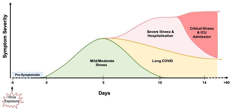
Fig. 4. COVID-19 symptomatology time course and outcomes. COVID-19 symptoms become apparent around 5 days after initial virus exposure. Symptoms peak, on average, 5 days later and, for the majority (>60%) of people, COVID-19 symptoms are mild to moderate in severity. Although symptoms subside for 4 in 5 of these mild/moderate cases after another 5 days, 1 in 5 people can experience persistent symptoms for 12 weeks or more (long COVID). Twenty percent of infected individuals require hospitalisation with breathing difficulties caused by viral pneumonia. Of these hospitalised patients, 1 in 5 develop ARDS and/or multi-organ failure requiring intensive care. Up to 40% of critical care patients can die. It has been estimated that around 17% of infected people never develop any symptoms of COVID-19 at any time.

Mild to Moderate COVID-19

The majority (> 60%) of infected individuals develop mild to moderate disease symptoms that generally resolve spontaneously after 6-10 days (Fig. 4.). However, around one in five of these individuals can have symptoms that persist from 4 weeks to over 12 weeks. Whether this post-COVID syndrome, generally known as long COVID, is a result of: long-term persistence of the virus; long term sequelae resulting from disruption of individuals’ immune and inflammatory responses; or due to something else entirely, is currently unknown.

Severe COVID-19

Around 20% of infected individuals require hospitalisation (Fig. 4.). Viral pneumonia, as evidenced by abnormal temporal lung changes on chest radiography, is the most common reason for admission, with patients displaying hypoxia due to a drop in blood oxygen levels. This leads to the majority of admitted patients suffering respiratory failure and requiring breathing support, which varies depending on individual patient need. Eighty percent of hospitalised patients are cared for in general medical wards.

Critical COVID-19

Twenty percent of hospitalised patients become critically ill and require invasive lung ventilation and support in high dependency and intensive care units (Fig. 4.). Sixty to eighty percent of these patients have ARDS and a hyper-inflammatory response. Unfortunately, up to 40% of critically ill COVID-19 patients die, although the death rate does vary with age. Severe COVID-19 can result in permanent damage and scarring to the lungs in survivors.

Multi-Organ COVID-19

Although the lungs are the primary site of COVID-19 pathology, COVID-19 is a multi-system disease exhibiting a range of complications resulting from damage to a number of different organ systems, including the heartbrainkidneyliver and vasculature (Fig. 3.). Elevations in levels of cardiac injury biomarkers are observed in 50% of patients with severe COVID-19, with a significant proportion of patients developing venous and arterial complications. Individuals with pre-existing endothelial cell dysfunction have an increased risk of damage caused by SARS-CoV-2 infection, which also increases hypercoagulability and results in a high incidence of venous thromboembolism and pulmonary embolism. Less commonly, SARS-CoV-2 infection can cause heart muscle inflammation and heart rhythm disturbances, such as atrial fibrillation. In cases of multi-organ dysfunction, specialist clinical support to reduce multi-organ failure and mortality is required.

COVID-19 PATHOGENESIS

The upper airways

The nasal cavity is the first site of infection (Fig. 5A.). Here, SARS-CoV-2 infects ciliated and secretory cells which express ACE2 and TMPRSS2. The viral load in the nasopharynx and oropharynx appears to be similar between infected people who stay asymptomatic and those who become symptomatic. There is little data at the current time relating to the extent to which SARS-CoV-2 spreads further in to the airways of asymptomatic people, but what data there is suggests the virus can spread deeper into the airways in some of these individuals and it is the host response which is a key determinant of outcome, including pre-existing immunity.

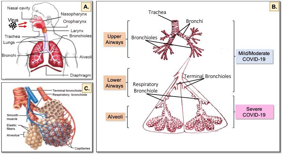
Fig. 5. The human respiratory tract and COVID-19. Inhaled virus particles bind to ACE2 receptors on the surface of epithelial cells in the nasopharynx and oropharynx (A). Disease severity is dependent on how deep SARS-CoV-2 virions progress into the airways. Restriction of virus infection to the upper and conducting airways (B) results in mild/moderate symptoms seen in >60% of infected individuals. Progression of the virus to the alveoli (C) affects exchange of oxygen and carbon dioxide between the lungs and the blood, which leads to the breathing difficulties seen in the 20% of patients hospitalised with severe COVID-19. In the 20% of hospitalised patients who become critically ill and require intensive care, damage to the endothelial cells of the capillary net encasing each alveolus leads to acute respiratory distress syndrome (ARDS) and/or multi-organ complications resulting from systemic effects of the virus.

Image sources: A. B. C.

Virus spreads from the nasal cavity to infect ciliated cells and non-ciliated mucus secreting (goblet) cells lining the bronchi (Fig. 5B.) as the virus progresses deeper into the lungs. During this phase, a vigorous innate immune response to clear the virus is triggered and the infected individual becomes symptomatic and feels unwell. Infection of lung epithelial cells triggers cell death and inflammatory responses. Keratin 5-expressing basal cells, which are the progenitor cells for the airway epithelium and are not infected by SARS-CoV-2, are induced to differentiate directly into ciliated cells so damaged epithelium can be repaired and recover from the infection. If the capacity for basal cell differentiation is impaired (e.g. due to excessive interferon lambda production), lung epithelial cell regeneration does not take place and airway damage increases, as happens in the lungs of patients with severe COVID-19.

The Lower Airways

Infection of club (Clara) cells located in the respiratory bronchioles in the lower airways facilitates spread of SARS-CoV-2 into the alveoli (Fig. 5C.), where oxygen and carbon dioxide exchange between the lungs and the blood is impaired in severe COVID-19.

Two types of epithelial cells line the alveoli (Fig. 6A.). Type I pneumocytes are large flat cells that are directly adjacent to the endothelial cells of the capillary network that closely encircles each alveolus (Fig. 5C.). Type I pneumocytes are critical for rapid exchange of oxygen and carbon dioxide between the blood and alveolar space. These cells are easily damaged in many forms of lung injury. Type II pneumocytes make and secrete pulmonary surfactant, which is required for effective gas exchange. Type II pneumocytes are also the progenitors of type I pneumocytes and are therefore important in the repair of alveolar damage. Both type I and type II pneumocytes help keep the alveoli free of fluid by contributing to active resorption of alveolar fluid and trans-epithelial ion movement. Type I and type II pneumocytes both express ACE 2.

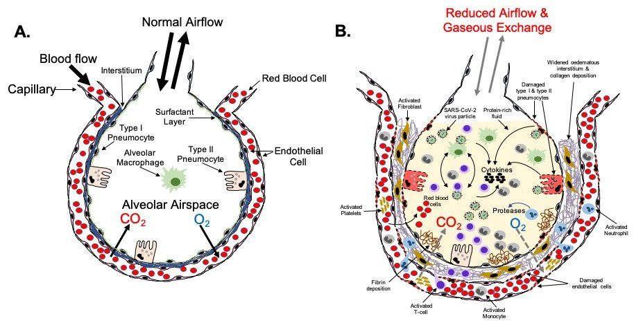
Fig.6. Severe COVID-19 lower respiratory tract pathophysiology. The alveolus (A) is the lung structure where gaseous exchange with the blood takes place. Oxygen in inhaled air and carbon dioxide in the blood diffuse out of, and in to, the alveolar space, respectively, via type I pneumocytes and the endothelial cells of the surrounding pulmonary capillary network. Surfactant secreted by type II pneumocytes aids this process by reducing surface tension at the air-liquid interface in the alveolus. Resident phagocytic alveolar macrophages provide a first line of defence against inhaled pathogens and noxious particles that make it down through the airways into the alveoli. In a healthy, non-diseased lung type I and type II pneumocytes keep the alveoli free of fluid. In severe COVID-19 (B), gaseous exchange  between the alveoli and blood is severely reduced. Damage to type I and type II cells by SARS-CoV-2 infection results in the alveolar space becoming filled with liquid containing dead cells, cell debris, proteins, invading immune cells, virus particles etc. In critical disease, extensive damage to the alveoli and endothelial cells of the surrounding capillaries, plus a hyperinflammatory response leads to ARDS and respiratory failure requiring prompt intensive care. In some patients this leads to death. Fibrotic changes resulting from disruption of the normal alveolar epithelial repair process lead to permanent structural changes to the lung micro-architecture that can have future health effects in some survivors depending on the scale and severity of these changes.

In addition to type I and II pneumocytes, there is a third cell type in the alveoli - alveolar macrophages (Fig. 6A.). These phagocytes are the first line of defence against infectious agents and noxious particles present in the environment that make it down through the airways to the alveoli. Like type I and type II pneumocytes, alveolar macrophages express ACE2.

Alveolar Inflammation

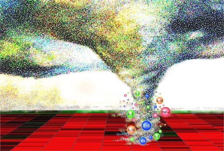
Type II pneumocytes express the highest levels of ACE2 and TMPRSS2 proteins and are believed to be the primary targets for SARS-CoV-2 infection in the alveoli. Type I pneumocytes and alveolar macrophages are also infected by SARS CoV-2. As the virus propagates in infected cells, released viral particles infect other type II pneumocytes and adjacent type I pneumocytes allowing spread of the virus to adjacent alveoli. Infected type II pneumocytes and alveolar macrophages secrete cytokines to recruit immune cells from the blood into the alveolar space to destroy virus-infected cells and extracellular virus. Uninfected neighbouring cells amplify this response by responding to, and secreting, additional cytokines. The innate immune response provoked in the pulmonary parenchyma is characterized by the recruitment of bone-marrow-derived monocytes to the alveolar space and their differentiation into alveolar macrophages.

The immunological hyper-response associated with COVID-19 ARDS appears to be a key driver of severity and death in patients. Although this hyper-inflammation is often referred to as a “cytokine storm” in a large number of publications, some investigators have questioned the relevance of this term in relation to COVID-19, given the cytokine levels actually measured in patients. Alveolar macrophages have a key role in initiating and propagating this hyper-inflammatory phenotype. Infected alveolar macrophages and activated T-cells recruited from the blood form a positive feedback loop that drives persistent alveolar inflammation. Neutrophils also appear to be important in the immune response and amplifying the damage seen in the lungs of patients with COVID-19 ARDS.

Alveolar Damage

Loss of type I and type II pneumocytes due to infection and subsequent cell death results in reduced gaseous exchange in diseased portions of the lungs (Fig. 6B.). Death of pneumocytes also results in focal alveolar flooding, causing the characteristic radiographic images seen in severe COVID-19. This is a result of a combination of factors including: reduced active resorption of alveolar fluid; reduced active transport of sodium from the alveolar space into the surrounding interstitium; damage to the endothelium of adjacent capillaries causing leakage of fibrinogen and other plasma proteins into the alveolar space; and formation of fibrin exudates. The flooding impairs the ability of pulmonary surfactant produced by type II pneumocytes to adsorb to the surface of the alveoli, thus increasing surface tension, and reducing oxygen exchange. As alveoli become blocked and collapse, appositional atelectasis occurs leading to loss of secondary lobules.

In most forms of lung injury, type II pneumocytes proliferate and differentiate into type I pneumocytes leading to restoration of the alveolar epithelium. However, the diffuse alveolar damage (DAD) seen in severe COVID-19 means that this repair process is disrupted because of depletion of type II cells. Activation of alternative pathways for epithelial repair in the most severe disease is likely to be the cause of the scarring and residual disease seen in some COVID-19 survivors. As type II pneumocytes inhibit fibroblast proliferation and the expression of extracellular matrix genes in fibroblasts, loss of these cells is likely to trigger fibrosis as fibroblasts migrate into the alveolar lumen. Imbalance in the renin-angiotensin system in COVID-19 may also favour lung fibrosis. The landscape and timescale of these changes will vary as new areas of the lung get infected and the innate and acquired immune systems respond.

Perivascular Damage

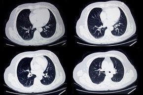
A key part of COVID-19 disease progression is damage to the pulmonary endothelial cells that are adjacent to the infected and damaged alveoli (Fig. 7). Analysis of lungs from patients who died of COVID-19 has highlighted several perivascular features, including: severe endothelial injury and disrupted endothelial cell membranes associated with intracellular SARS-CoV-2 virus; widespread vascular thrombosis with microangiopathy and occlusion of alveolar capillaries; and significant new vessel growth through intussusceptive angiogenesis. Changes in the pulmonary vasculature on chest computed tomography (CT) appear to be predictive of adverse clinical outcomes in COVID-19 patients.

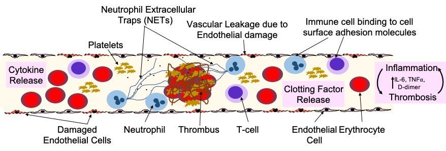
Fig. 7. Endothelial damage and dysfunction in COVID-19 lung pathology and extra-pulmonary organ damage. Endothelial dysfunction is associated with the development of severe COVID-19 and life-threatening extra-pulmonary complications. Endothelial cells can be damaged and activated by: direct viral infection; by pathogen-associated molecular patterns (PAMPs) derived from SARS-CoV-2; damage-associated molecular patterns (DAMPs) from dead or dying endothelial and immune cells; or pro-inflammatory cytokines (e.g. interleukin 6/IL-6 and tumour necrosis factor alpha/TNFa). Endothelial dysfunction and injury lead to: increased vascular permeability; increased endothelial inflammatory response; reduced levels of nitric oxide, causing vasoconstriction; and impaired angiogenesis. Activated endothelial cells release cytokines that attract immune cells that bind to adhesion molecules expressed on the surface of the endothelial cells and also produce cytokines. This stimulation of the immune response leads to hyper-inflammation, which leads to further endothelial damage. Release of clotting factors (Von Willebrand Factorthrombinfibrin and tissue factor), platelet activation and NETosis of neutrophils, drives thrombus formation and increases in the levels of D-dimer. Neutrophils also amplify  endothelial damage by releasing a number of proteolytic enzymes and reactive oxidant species that damage cells.

Pulmonary endothelial cells (ECs) have a key role in optimising gas exchange, controlling barrier integrity and function, and regulating pulmonary vascular tone in health and disease. They are involved in most lung diseases either as a direct participant, or as a victim of collateral damage. Importantly, endothelial cells express ACE2, so SARS-CoV-2 may alter vascular homeostasis by directly infecting endothelial cells. Analysis of patient tissue samples and organoids show that SARS-CoV-2 can infect endothelial cells, even though cultured primary endothelial cells are resistant to infection.

Alternatively, instead of direct infection by SARS-CoV-2, vascular damage in COVID-19 may be driven by hypoxia, hyper-inflammation and immune dysregulation. Platelet-neutrophil communication and activation of macrophages can induce a range of pro-inflammatory effects, including: cytokine release; the formation of neutrophil extracellular traps (NETs); increasing fibrin generation; and inducing microthrombus formation in the microvasculature. NETs can damage the endothelium and activate both extrinsic and intrinsic coagulation pathways and appear to have a significant role in COVID-19 disease severity.

EXTRAPULMONARY ORGAN DAMAGE

As noted earlier, in addition to lung pathology, SARS-CoV-2 infection can result in a number of extrapulmonary complications involving the haematological, cardiovascular, renal, gastrointestinal, hepatobiliary, endocrinologic, neurologic, opthalmologic and dermatologic systems (Fig. 3.). Potential mechanisms underlying the pathophysiology of this multi-organ injury include: direct viral infection of organs; endothelial cell damage and thromboinflammation; immune response dysregulation; and dysregulation of the renin-angiotensin-aldosterone system (RAAS). The role that each of these mechanisms plays in extra-pulmonary organ damage in COVID-19 is unclear at the present time. However, the pathophysiology underlying the systemic effects of COVID-19 is likely to be multi-factorial, with different mechanisms predominating in different patients.

Direct Infection

The presence of ACE2 and TMPRSS2 in a range of different extra-pulmonary organs makes direct infection a plausible mechanism for multi-organ injury in COVID-19. Evidence for direct viral infection of the kidney and other organs in patients with COVID-19 and the detection of viral RNA in blood and urine samples from patients supports the idea of systemic spread of the virus. However, the mechanism(s) of extrapulmonary virus spread have yet to be elucidated.

Endothelial Cell Dysfunction

Endothelial cells have a number of physiological functions and are involved in a range of different diseases. Because they line the inside surface of blood vessels, they traverse a wide range of organ systems. Endothelial damage and endothelialitis are found in the vascular beds of a number of organs in patients with COVID-19 and this can trigger thromboinflammation leading to microthrombi deposition and microvascular dysfunction. Recent work suggests that inflammation and vascular damage may be the primary causes of neurological symptoms in COVID-19 patients. As a result, COVID-19 is increasingly being seen as an endothelial disease, with endothelial cell dysfunction (Fig. 7.) being the common link between many of the different complications seen in COVID-19 patients, from multi-organ damage, to blood clots and stroke. Moreover, pre-existing endothelial dysfunction is the common denominator among co-morbidities such as hypertension, diabetes, obesity, cardiovascular disease and ageing in individuals with an increased risk of severe COVID-19. Published data suggests that, at least in some COVID-19 patients, acute multi-organ thromboembolism may precede, or present disproportionately, over respiratory involvement. Organ dysfunction is also associated with excessive NET formation and vascular damage.

Immune Dysregulation

As mentioned previously, the host immune response is important in governing individual patient outcomes in COVID-19, whether that is a reduced response, or an over-enthusiastic response by the innate immune system. Markers of inflammation are predictive of critical illness and mortality in COVID-19 patients, supporting the idea that hyperinflammation is a driver of multi-organ failure in COVID-19. The positive effects of dexamethasone and IL6 receptor antagonist therapy also indicate the importance of the immune response in disease progression and severe pathology.

Renin-Angiotensin-Aldosterone System (RAAS) Dysfunction

ACE2 is a key regulator of RAAS in cardiovascular and pulmonary homeostasis. Vascular damage-induced by SARS-CoV-2, combined with pre-existing endothelial dysfunction caused by hypertension, diabetes, obesity, or age appears to be a contributory factor in COVID-19 related morbidity and mortality, including the vasculopathy and coagulopathy seen in COVID-19. Therefore, dysregulation of the RAAS offers another plausible mechanism for SARS-CoV-2 tissue damage of extra-pulmonary organs.

COVID-19 VACCINES AND THERAPEUTICS

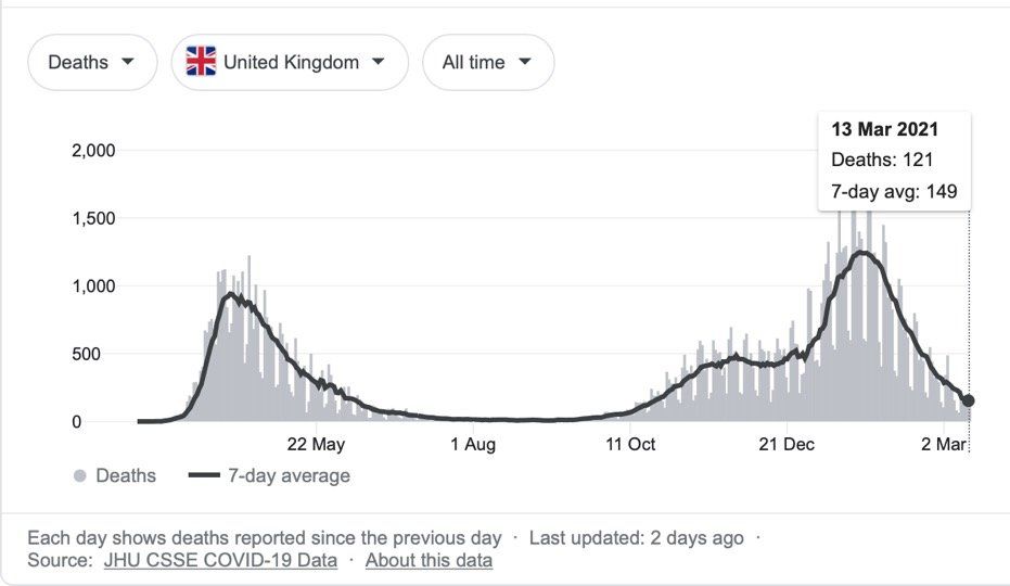
As of  12th March 2021, there have been 118,742,439 COVID-19 cases worldwide, resulting in 2,632,955 deaths. In the UK there have been 4,241,677 cases and 125,168 deaths, although the number of cases and deaths has declined markedly from the late January 2021 peak, as a result of lockdown measures and an efficient vaccination programme.

Twelve vaccines have been approved at the time of writing. Thanks to the rapid availability of the SARS-CoV-2 genome sequence and advances in vaccine technology, the speed of COVID-19 vaccine development has been remarkable, taking less than a year from genome sequence determination to the UK approving the Pfizer-BioNtech vaccine, quickly followed by approval of the AZ-Oxford vaccine. This was a timescale previously unimaginable using traditional vaccine development methodology.

As of 10th March 2021, 23,053,716 people, representing 34.59% of the UK population have received at least one dose of the Pfizer-BioNtech or AZ-Oxford vaccine. A smaller number, 1,351,515 people, representing 2.03% of the UK population, have been fully vaccinated after receiving two doses. The number continues to grow daily.

The roll out of SARS-CoV-2 vaccines is already having a major impact on the incidence of severe COVID-19. The emergence of new SARS-CoV-2 variants that have increased rates of transmission and/or may reduce or abolish the effectiveness of the current vaccines is a concern. However, global surveillance of virus variants by sequencing, combined with the ability to quickly develop new vaccines, will reduce this threat. As the risk of new variants is lowered if the rate of new infections (and viral replication) is reduced, equitable access to vaccines for the world’s population, particularly poorer countries, will also be very important in reducing the number of new variants in the medium to long-term.

Can SARS-CoV-2 be eradicated, or will we just have to learn to live with it? I suspect the latter is the more achievable scenario, at least in the medium term, given: the level of vaccine hesitancy that exists; the proportion of people in the world who have still to be vaccinated; and the continuing emergence of virus variants.

The global research effort focussed on SARS-CoV-2 and COVID-19 has been unprecedented. In the time it has taken to write this article, over 8,000 new papers have been added to the 103,276 papers that existed in PubMed when I started writing! However, in contrast to the situation with vaccines, only modest progress has been made with regard to the development of anti-viral drugs to prevent SARS-CoV-2 infections, and other drugs to stop COVID-19 disease from worsening. This lack of success is not due to lack of effort, just a reflection of the scale of the problem that COVID-19 has presented to the scientific and medical worlds. In truth, drug discovery has still to reach the level of speed and refinement that vaccine technology currently has. So far, remdesivir is the only anti-viral drug approved for treating Covid-19, but its use is limited to clinical settings and, at best, it appears to be only modestly effective. Emergency use applications (EUAs) have also been issued by the US Federal Drugs Administration (FDA) for several unapproved monoclonal antibody therapies to treat mild to moderate COVID-19 in children and adults. In terms of immunomodulators for severe COVID-19 disease, dexamethasone and tocilizumab, particularly when used together, have been shown to reduce COVID-19 mortality.

How can the world be better prepared for similar threats in the future? Preparation for the next crisis occurs needs to begin now. Pre-emptive development of effective and readily available drugs that can be quickly tested in new diseases as they arise is required, mirroring the pre-emptive strategies that allowed the rapid development of SARS-CoV-2 vaccines. The last point notwithstanding, there will always be a lag between the outbreak of a new pandemic and the delivery of an effective vaccine, so antiviral drugs will be the primary tools that can be used to help keep populations safe during this period. There will also be a need for immunomodulatory drugs to treat severe disease in susceptible individuals and reduce mortality. In the main, I believe this can be most effectively approached using drug re-purposing strategies, rather than starting drug discovery programmes from scratch. This is an opportunity for pharmaceutical companies to work together with government agencies and academia in public-private consortia to pool resources and expertise to form a centralised library of drug compounds that can be rapidly tested in clinical trials. Furthermore, investment in identifying future primary threats and making a societal commitment to pre-emptive preparation is also needed. This will require a high degree of coordination and the involvement of scientists from fundamental researchers, to drug development experts. Given the remarkable response to the current pandemic, this is achievable. Ideally, the work needs to be undertaken globally, to spread the cost and maximise expertise and resources, as well as ensure that important research on other diseases is not disadvantaged.

What about the use of viral challenge studies in the development of new anti-viral drugs and vaccines? Approval of the world’s first challenge study in the UK is a key step towards this goal. If such studies can be conducted safely on healthy volunteers they will be important in facilitating the development of new vaccines and therapeutics. However, they will not be of value in the development of drugs to target severe disease as it would clearly be unethical to deliberately trigger life threatening symptoms in volunteers. For this, standard clinical trials using patients hospitalised with severe disease will still be required, underlining the importance of having libraries of immunomodulatory drugs ready for rapid testing in such studies when a pandemic arises.

CONCLUSION

Although the scientific and medical response to the COVID-19 pandemic has been exceptional, and the progress made in understanding the disease and developing vaccines has been astounding, the social and economic cost underlines the importance of learning from the current pandemic and being better prepared for the next one. Clearly, it is not a question of if, but when, it will occur again and how well the world will be prepared for it next time…

by Paul Whittaker 9 June 2025
Modified from Image Source Animal studies in sheep and mice , as well as evidence from trials in humans and case studies of compassionate clinical use, indicate that phage therapy is efficacious, safe , and non-toxic . A 2022 systematic review of clinical data obtained from phage therapy clinical trials, safety trials, and case studies between 2000 and 2021 for difficult to treat bacterial infections in several medical disciplines, concluded that phage therapy given via different routes of administration is well tolerated and safe with a low incidence of side effects. Unfortunately, heterogeneity between different clinical studies precluded a meta-analysis of the data, highlighting the need for high quality clinical trials to improve knowledge on long-term patient and disease outcomes. The use of purified phage to treat superficial bacterial infections appears to be efficacious safe, and side effect free, even when delivered by invasive routes of administration (e.g. intravenous and intra-articular ), or used in immunocompromised patients. Some would argue that phage therapy appears to have a better safety profile than antibiotics , and has a minimal impact on commensal flora , thus reducing the likelihood of opportunistic infections. Consistent with our natural exposure to phages, there are no reports of allergic responses to phages, potentially making them suitable alternatives for patients with antibiotic hypersensitivity. Because phages can kill bacterial cells quickly , release of endotoxins from lysed bacterial cells in severe infections is a potential safety concern. However, it has been demonstrated that phage lysis releases less endotoxin than beta-lactam antibiotics. Another potential issue with phage therapy is that phage cocktails might have effects on non-targeted bacteria and so affect the human microbiome. However, phage therapy does not appear to have an adverse effect on gut microbiota and has less of an effect on gut diversity than antibiotics, having a beneficial effect on gut health. Studies have suggested that bacteriophages can interact with eukaryotic cells, significantly influencing the functions of tissues, organs, and systems of mammals, including humans. In addition, there are concerns regarding the long-term impact of phages on the human immune system. In terms of effects on innate immunity , phages appear to induce anti-inflammatory responses. In terms of effects on the adaptive immune response , phages can be immunogenic, but are not very effective at inducing a specific immune response. With regards to phages inducing anaphylaxis , no cases have been reported. Autoimmune responses to phage therapy are a possibility, however, their immunomodulatory role, particularly in curbing inflammation means that phage therapy is being explored for the treatment of autoimmune liver diseases . Although the indications are that phage therapy is safe, with few adverse effects, current research on phage safety monitoring lacks sufficient and consistent data for regulatory purposes, which would require a standardized phage safety assessment to ensure a robust evaluation of the safety profile of phage therapy. Although the Australian STAMP protocol is not a randomised clinical trial, it does provide a framework for the collection of higher quality efficacy and safety data than individual case studies.
by Paul Whittaker 8 June 2025
Image Source In this second article about phage therapy, I will be focussing on its use in different countries, with special emphasis on the UK. Phage Therapy in the UK In the UK, phages are classed as a biological medicine and none are licensed for clinical use. As a result, phage therapy is applied on a compassionate use basis as an unlicensed medicinal product (a “ special ”). Phages imported for use as an unlicensed medicine in the UK do not need to be manufactured according to Good Manufacturing Practice ( GMP ), however, the Medicines and Healthcare products Regulatory Agency ( MHRA ) must be notified at least 28 days prior to importation, and doses imported are limited to a small number. Paradoxically, phages manufactured in the UK must be produced to GMP, including phage for use in clinical trials. As a result, the current clinical provision of phage therapy in the UK is ad hoc and relies heavily upon networking with international sources of phages , including organisations such as Phage Directory , who help connect clinicians who want phages for clinical use, with groups who have appropriate phages. The MHRA recently published a document on the regulatory considerations for therapeutic use of bacteriophages in the UK. According to Phage-UK there have been 24 clinical trials involving phage therapy since 2020. Phage Therapy in Other Countries Globally, different countries have different regulatory frameworks for the clinical use of phage therapy. Eastern European countries have over 100 years of phage therapy experience. Russia and Ukraine allow open use and commercialisation of phage products. Phages are a standard medical application in Georgia . In Poland , specialised institutes have supplied personalised phage products to physicians since 2005. In the European Union (EU) , phage therapy is not approved as a standard medicinal product for human use. Like the UK, It is primarily used in compassionate use cases, clinical trials, or for individual experimental therapy attempts. Although a European Medicines Agency (EMA) guideline exists for veterinary bacteriophage medicinal products , there is currently no corresponding regulatory guidance for human use of such products in the EU. The EMA opened a public consultation on a concept paper on the development and manufacture guidelines for human bacteriophage medicinal products tailored to phage therapy on 23 rd December 2023, which ended on 31 st March 2024. In the meantime, a regulatory roadmap for phage therapy under EU pharmaceutical legislation has been published . Belgium has implemented a phage therapy framework focusing on magistral phage preparations that allows patients to actively seek access to personalized phage therapy. In the magistral approach , individual phages are prepared according to a phage monograph (a standardized document that provides detailed information about a specific bacteriophage, its properties, and its potential use in phage therapy), and reference laboratories provide quality control (QC) testing. Clinicians prescribe phage cocktail preparations for use in specific patients, which are then prepared by pharmacists. Based on the Belgian model, a general chapter on phage therapy medicinal products was published in 2024 by the European Pharmacopeia . In Australia , all clinicians and researchers within the Phage Australia network have adopted the Standardised Treatment and Monitoring Protocol for Adults and Paediatric Patients ( STAMP ). STAMP is a clinical protocol for administering and monitoring phage therapy, rather than the phage product. As a result, STAMP looks at the process, not the product and means that different patients can be treated with different phages at different sites of infection, but the treatment protocol is standardised. The STAMP protocol has been approved by Australia's national ethics committee and endorsed by Australia's national infectious disease physician society, as well as its paediatric arm. In the US , phage therapy is not yet an FDA licensed treatment. However, it available under a special programme called the “ expanded access eIND system ” . For patients who have exhausted standard-of-care therapy, an application is submitted to the FDA by the treating physician, where a patient meets a list of criteria. UC San Diego's IPATH is the first dedicated phage therapy centre in North America. They focus on treating patients with life-threatening multi-drug resistant infections through the FDA's compassionate use program. IPATH also works to advance phage therapy into clinical trials and provides guidance to physicians worldwide on phage therapy protocols. The PhagesDB database details over 25,000 new phages identified by the SEA-PHAGES programme at the University of Pittsburgh. One particular phage from this collection, called Muddy, has been used therapeutically in a cystic fibrosis patient infected with a multi-drug resistant strain of Mycobacterium abscessus. Creative Biolabs is developing libraries of characterised phages, as sell as platforms for identifying and then producing phages to GMP standard for formulation and delivery. Israel focusses on compassionate use of phage therapy, with the Israeli Phage Therapy Center conducting all of the steps required, from phage isolation and characterization to treatments for non-resolving bacterial infections. In India , phage therapy is offered as compassionate Phage Therapy regulated by the Declaration of Helsinki and coordinated by the Central Drugs Standard Control Organization. Vitalis Phage Therapy has created a framework for patients to access phage therapy in India. In China , there are two routes to using phage therapy applications. Phage products with fixed ingredients are regulated as innovative biological products . Personalized phage therapies, on the other hand, need to go through investigator-initiated trials (IIT) and, if successful, the phage therapeutic can then be used at certain institutions. Expanding the Use of Phage Therapy in the UK To progress past the ad hoc use of phage therapy in the UK, the infrastructure to support the route from patient enrolment, through isolation and identification of pathogenic bacteria and therapeutic phages, to formulation, administration and monitoring of efficacy, as well as phage resistance, will need to be put in place. A key requirement for clinicians in the UK will be the ability to access phages that are efficient at killing the strain of bacteria causing an infection. A roadmap for the delivery of clinical phage therapy to the UK has been proposed, which would require: expansion of existing phage biobanks; the development of both personalised treatments for individual patients; and off-the-shelf phage cocktails that could be used to treat large numbers of patients. Innovate UK has developed the Phage Innovation Network to help drive the use of phage based therapy in the UK and build on the expertise of the many phage experts based in the UK. To enable this, systematic libraries of a diverse array of phages that are well characterised and curated, as well as manufactured to GMP standard will be needed. The Citizen Phage group at Exeter University , uses volunteer citizen scientists to collect samples from a wide range of environments to facilitate the laboratory identification of new phages for therapeutic use. The UK Phage Library at the University of Leicester is aiming to develop libraries of phages which can be screened against specific bacterial strains to identify phages they are sensitive to. Unfortunately, these phages cannot be used in patients in the UK due to lack of GMP phage manufacturing capability, but they are provided for use in other countries whose regulatory frameworks permit their use. On the commercial side, Nexabiome in Glasgow aims to provide an end-to-end service covering phage identification and isolation, to production and formulation. Establishing a UK Phage Manufacturing Facility that can produce phage preparations for both commercial and non-commercial customers that are suitable for administering to patients would be a key requirement for widening the use of phage therapy in the UK. Towards this end, UK Phage Therapy is working with public and private partners to establish a centralised phage susceptibility testing and GMP phage production facility in the UK via the Centre for Process Innovation (CPI) . The Centre for Phage Research is also working with regulators, policymakers and other stakeholders to establish frameworks and pathways to enable public access to phage products. Whether the UK will remove the requirement for GMP manufacture of phages for Phase I clinical trials, as is the case in the US, is unclear. However, phase II/III trials would still require GMP manufacture for off-the-shelf phage products. Phage-UK has developed a standardised protocol for the treatment and monitoring of phage therapy in UK patients suffering from cystic fibrosis and bronchiectasis , where there is no alternative treatment available. The protocol, based upon the Australian STAMP protocol, is a document that assists clinicians making a submission to their NHS Hospital Board for approval to use phage therapy on a named patient basis. Presumably, this protocol could be adapted for the treatment of UK patients with other serious infections such as urinary tract infections (UTIs), prosthetic joint infections (PJIs) and sepsis. A benefit of using such a standardised protocol would be the collection of safety and efficacy data on phage therapy that could then inform the design of subsequent clinical trials of phage therapy in the UK. Conclusion For most countries, compassionate use is the major pathway for patients to access phage therapy. Belgium (with the magistral approach), and Australia (with STAMP), currently lead the way in developing frameworks to facilitate patient access to phage therapy. The lack of such frameworks in other countries, including the UK, reflect the view that phage therapy is still an experimental treatment that requires more convincing clinical evidence of efficacy. In the UK, it will be interesting to see if any of the recommendations made in the March 2024 Policy Paper on the antimicrobial potential of bacteriophages, published under the Conservative government in power at the time, are followed up by the current Labour government.
by Paul Whittaker 3 June 2025
Image Source: The growing global problem of antibiotic resistance , and the lack of new antibiotics being developed, has rekindled an interest in phage therapy: the use of viruses (bacteriophages) that specifically target and kill bacteria, as a treatment for antibiotic resistant infections in humans. This article is an overview of phage therapy and is the first in a series where I will explore aspects of this technology in greater detail. Links to papers and websites that contain diagrams or graphics relevant to this article are provided at the end of this article, as are links to PubMed search results for phage therapy papers, reviews and clinical trials. Bacteriophages In 1986, nanotechnology pioneer K. Eric Drexler imagined a dystopia where invisible self-replicating nanobots proliferated voraciously and took over the entire planet. Spooked by Drexler’s nightmare, Prince Charles (the heir to the British throne at the time, but now King Charles III) requested that the eminent Royal Society investigate the risks that nanotechnology posed. However, the reality is that nano-scale self-replicating voracious killers have existed on earth for over 4 billion years. They can make hundreds of copies of themselves in as little as 15 minutes, and are found in vast numbers everywhere on our planet. Thankfully, they are not harmful to humans. Instead, they infect and destroy bacteria in a process perfected over the eons. Known as bacteriophages , these biological entities were discovered at the beginning of the twentieth century. Bacteriophages (usually referred to as phages - derived from the Greek word “phagein”, meaning “to devour/eat”) are viruses composed of a nucleic acid genome encased in a phage-encoded protein capsid shell. Phages are found in three basic structural forms : icosahedral head with a tail; icosahedral head without a tail; and filamentous. They infect and kill bacteria by replicating inside bacterial cells, then breaking open ( lysing ) the infected cells before releasing large numbers of new progeny phage particles. In the laboratory, this process is visualised and monitored using plaque assays . Phages are ubiquitous and diverse . It has been estimated that there are 10 31 bacteriophages on the planet, more than every other organism on Earth, including bacteria. Phages can be isolated from sources where high numbers of bacteria occur, such as human sewage , soil , rivers , faeces , even slime in a stream . Phages are specific to individual bacterial species and strains and do not infect mammalian cells. However, because of the microbiome , the human body contains large numbers of phage particles and varieties of phages (the so called phageome ) and, as a result, phage can interact with mammalian cells. Phages are fascinating biological entities. As an undergraduate biology student I learned of the importance of phages as key experimental tools in the development of the fields of molecular genetics and molecular biology. As a post-graduate biochemistry student I worked with phages in the lab of the late Pauline Meadow , who used them as a way of identifying lipopolysaccharide (LPS)-defective mutants of Pseudomonas aeruginosa. As a doctoral student in molecular biology working on DNA methylation in the slime mould Physarum polycephalum, and as a postdoctoral researcher working on human genome analysis, I used phage lambda cloning vectors to construct genomic DNA libraries for gene isolation (e.g. tubulin genes from the parasite Trypanosoma brucei) and for the physical mapping of human genomic DNA (e.g. the human dystrophin-encoding gene using a specially modified phage lambda vector I developed). The Challenge of Bacterial Anti-Microbial Resistance More widely referred to as anti-microbial resistance , or AMR , bacterial AMR (bAMR) occurs when bacteria develop the ability to defeat the antibiotics designed to kill them. This resistance can result from several different mechanisms . I n this article I am referring specifically to bAMR, and not viral, fungal, or parasitic AMR . A systematic analysis published in 2022 estimated that there were 4.95 million deaths associated with bAMR globally in 2019. As a result, bAMR has the potential to affect each and every one of us by impacting the treatment of illnesses, surgical procedures and cancer treatment, as well as increasing rates of death. The increase in prevalence of bAMR has led to fears of future pandemics caused by drug-resistant bacteria. In the UK, the Government and the National Health Service (NHS) have both developed action plans to tackle bAMR which emphasise optimising and reducing exposure to antibiotics. Despite these measures, however, new antibiotics and alternatives to antibiotics are still needed, particularly in cases where infections are refractory to antibiotic use. Developing new classes of antibiotics is challenging . The greater portion of recently approved antibiotics have tended to be derivatives of existing classes of drug compounds. Although pharmaceutical giant Roche recently reported the identification of a promising new class of antibiotic molecules that target carbapenem-resistant Acinetobacter baumannii (CRAB), many big pharma companies have ceased antibiotic development. As a result, small biotech companies are leading research and development efforts in this area. Phage Therapy Case studies in the scientific literature , and success stories described in the press and in books have highlighted the use of phage therapy to treat infections caused by antibiotic-resistant bacterial strains. However, phage therapy as a way of treating bacterial infections is not new. Phages were first used to treat bacterial infections in 1919 (the “pre-antibiotic era”), but the approach never really gained traction in the West, particularly after penicillin was discovered in 1928 and became the favoured way to treat bacterial infections in the 1940s. Despite this, phages have continued to be used in Russia , Georgia and Poland as an alternative to antibiotics since the early 20 th century . The excellent book “The Good Virus” by Tom Ireland, gives a vivid and detailed account of the history of phage therapy and how interest in it as an approach to treating bacterial infections has waxed and waned over the past century. Now, because of the growing threat of bAMR, there has been a resurgence of interest in using phages to tackle antibiotic resistant infections. As some phages degrade biofilms, phage therapy also potentially provides a way to deal with the antibiotic tolerance seen in some chronic diseases resulting from biofilm production (e.g. cystic fibrosis ). Also of interest is the potential to use phage resistance as a way to steer bacteria towards an antibiotic-sensitive phenotype. Unfortunately, in many parts of the world current regulations restrict the application of phage therapy to individual 'compassionate use ' in patients with infections where antibiotics have failed. In the UK phage therapy has been used sparingly to treat Pseudomonas and Mycobacterial infections mainly due to the lack of sustainable access to phages manufactured to good manufacturing practice ( GMP ) standard. The first successful clinical trial of phage therapy in the UK was published in 2009. Since then, phage therapy has been used in the treatment of patients with diabetic foot ulcers and cystic fibrosis . There has often been a difference between the results of individual real world case reports of successful phage therapy and the results of larger scale studies. Therefore, high quality clinical trials of phage therapy in the treatment of a range of conditions are needed to provide a solid evidence base on the efficacy and safety of phage therapy in human patients to support wider clinical use. A retrospective observational analysis of 100 consecutive cases of personalised phage therapy carried out by a Belgian consortium using combinations of 26 bacteriophages and 6 defined bacteriophage cocktails reported clinical improvement and eradication of targeted bacteria for 77.2% and 61.3% of infections, respectively. However, eradication was 70% less when antibiotics were not used concurrently. Recently, there have been calls for the British Government to invest in phage therapy as a way to tackle bAMR. As a first step, a report published in January 2024, following a Parliamentary Inquiry in 2023, recommended that the British Government bring together phage experts and stakeholders (scientific, clinical and regulatory) to assess what would be required to enable phage therapy to be used more widely in the National Health Service (NHS) and other UK healthcare settings. A Government response to this report which supports these recommendations and makes 18 additional recommendations across 4 themes, has now been published . The Innovate UK Phage Knowledge Transfer Network has been established to provide a forum for funders and phage researchers to discuss these matters and ways forward, including multi-party collaborations and co-investments by public and/or private funders. Discussion The dramatic results seen in sick people who have received phage therapy as a last ditch treatment when conventional antibiotic therapy has failed, provides a compelling narrative for its potential in the treatment of bAMR. However, the body of evidence required to convince regulatory authorities, governments and the medical establishment of phage therapy efficacy is clearly lacking at the moment. Even if that data is forthcoming, it is unlikely that phage therapy will ever replace antibiotics. More likely, phage therapy will continue to be used in a personalised way to treat infections that are resistant to standard antibiotic therapy, but in the future it is reasonable to envisage clinical scenarios where phages might be used in conjunction with antibiotics. The judicious use of phages might help protect and preserve existing antibiotics and combining the two appears to be more effective than either on their own. Phage therapy may also be useful for treating people who are allergic to antibiotics and may not have other treatment options. It is, of course, entirely possible that phage therapy for humans never progresses past the stage it is at now. Maybe new classes of antibiotics will be discovered. Maybe other antimicrobial therapeutic modalities will be invented, or discovered. Hopefully, though, the many initiatives taking place worldwide will result in the development of a safe and clinically proven version of phage therapy that will become part of an expanded therapeutic toolkit. However, what is clear at present is that while these challenges are being tackled, phages are already being deployed in various animal and non-medical scenarios such as food safety and environmental pathogen control (e.g. aquaculture ). As I explored the extensive literature around phage therapy , I realized that I could only scrape the surface of this subject in such a short article. But it is a fascinating field with therapeutic potential. Therefore, in later blog articles, I will discuss aspects of phage therapy; such as safety, phage production, commercialisation, and the regulatory approaches to using phage therapy in different countries round the world, including the UK, in more detail. Links to websites and papers with relevant diagrams and graphics: Structure of a bacteriophage Bacteriophage life cycle Phage lambda structure How bacteriophages infect and lyse bacterial cells Phage therapy Bacteriophage plaque assay Overview of antimicrobials Antibiotic Resistance Causes of antibiotic resistance Antimicrobial resistance worldwide PubMed literature links: Bacteriophage papers Bacteriophage therapy papers Bacteriophage therapy reviews Bacteriophage therapy randomised controlled trials Phage therapy systematic reviews
by Paul Whittaker 28 May 2025
The importance of lung structural abnormalities
by Paul Whittaker 20 May 2025
Targeting p38MAPkinase
by Paul Whittaker 18 April 2021
Image Source.
by Paul Whittaker 25 March 2021
Image Source
by Paul Whittaker 4 May 2020
A collection of links to information sources relevant to the coronavirus pandemic. From disease and healthcare information, to drug therapies and diagnostics, as well as epidemiology and clinical trials in progress. Association of British Pharmaceutical Industries Up-to-date information on what the pharmaceutical industry is doing to tackle the outbreak Diagnostics webinar slides British Broadcasting Corporation BBC Horizon programme investigating the scientific facts and figures behind the Coronavirus pandemic as at 9th April 2020 British Medical Association COVID-19 Guidance Official guidance for doctors British Medical Journal Coronavirus Hub Support for health professionals and researchers with practical guidance, latest news, comment and research British Society for Antimicrobial Chemotherapy Information and the latest guidance from health bodies, academic publications and other professional societies British Thoracic Society COVID-19 Information Information, guidance and resources to support the respiratory community Centers for Disease Control and Prevention Coronavirus disease portal - up to date genomics and precision health information on coronavirus US government public health information Cochrane Library COVID-19 Information Cochrane Reviews and related content from the Cochrane Library relating to the COVID-19 pandemic COVID-19 Diagnostics Resources Diagnostics resource centre is designed to support policymakers and healthcare providers with up-to-date information on tests and testing for SARS-CoV-2 Resource page to facilitate information sharing and provide technical expertise to teams developing COVID-19 tests Drug Target Review Latest news and updates relating to COVID-19 drug discovery efforts European Respiratory Society COVID-19 Resource Centre European Respiratory Society (ERS) and European Lung Foundation (ELF) resources on SARS-CoV-2 and COVID-19 as it is published International Severe Acute respiratory and Emerging Infection Consortium (ISARIC) COVID-19 Resources COVID-19 clinical research resources Medical Research Centre for Epidemiological Analysis and Modelling of Infectious Diseases All output from the Imperial College COVID-19 Response Team, including publicly published online reports, planning tools, scientific resources, publications and video updates Public Health England PHE COVID-19 dashboard Royal College of Pathologists Latest information available on COVID-19 relevant to the practice of pathology Royal Society of Biology How the RSB is responding to the COVID-19 pandemic, by area of activity Bulletin covering the latest news and science behind the outbreak and the response Royal Society of Medicine Roundup of official guidance and information, tools and helpful resources on COVID-19 for healthcare professionals Resources on COVID-19 national trials University of Oxford Centre for Evidence-Based Medicine Rapid reviews of primary care questions relating to the coronavirus pandemic, updated regularly US National Institutes of Health Latest research information from NIH Wiley Online Library Articles related to the current COVID-19 outbreak, as well as a collection of journal articles and book chapters on coronavirus research freely available to the global scientific community
by Paul Whittaker 1 May 2020
Image Source “ By the help of microscopes, there is nothing so small, as to escape our inquiry; hence there is a new visible world discovered to the understanding. ” Robert Hooke , 1635 - 1703. The history of microscopes is one of ever higher magnification, enabling scientists to pry deeper and deeper into the minute details of nature invisible to the naked eye. Now microscopy is playing a key role in visualising the molecular architecture of proteins and macromolecular complexes; information which is critical to understanding biomolecular function and discovering new drugs. X-ray crystallography has been the most widely used technique for obtaining atomic level detail, but it does require a large amount of sample and the generation of crystals, which can be problematic. Nuclear magnetic resonance (NMR) does not require crystallization, but it does require large amounts of sample and isotopic enrichment and the technique has generally been restricted to small proteins, single protein domains, or small RNAs. Single-particle cryo-electron microscopy (cryo-EM) is a structural biology technique, first developed back in the 1970s, which does not require crystallization, or large amounts of sample. Using a flash-freezing process that fixes proteins in thin films of ice, cryo-EM avoids the problems of crystallization. Then, thousands of 2-D images of proteins caught in random orientations are stitched together to reveal the 3D structure. Advances in detector technology and software algorithms now mean that cryo-EM can be used for the determination of biomolecular structures at near-atomic resolution , including proteins and macromolecular assemblies “intractable” to X-ray crystallography and NMR. In an application relevant to the current COVID-19 pandemic, cryo-EM has recently been used to study the functional evolution of coronavirus spike proteins and provide a structural basis for the inhibition of coronavirus replication by Remdesivir . Cryo-EM is now being used for a range of applications from studying eukaryotic DNA replication, to structure-based drug discovery (SBDD), as showcased during a recent ELRIG webinar on this topic, held on 2nd April 2020 entitled “From Blob-ology to Near Atomic Resolution Structures: Current Uses of Cryo-EM within Biological Research”. While the number of protein structures being determined by cryo-EM is booming , the cost of a microscope (up to £5/$7 million), the need for custom laboratory facilities and the associated operational costs, means that many structural biologists have no access to this technology. Jason Van-Rooyen explained how The Electron Bio-Imaging Centre ( eBIC ) at Diamond in Oxfordshire provides state-of-the-art cryo-EM equipment and expertise, as well as molecular and cellular cryo-electron tomography for use by academic and industrial scientific groups . Use of the eBIC cryo-EM facility has resulted in 113 publications at the time of writing this article, ranging from the determination of cryo-EM structures of the Escherichia coli RecBCD complex to the cryo–EM structure of RagA/RagC in complex with mTORC1 . Planned, are the provision of a Dual Beam Scanning Electron Microscope (SEM) and Focused Ion Beam (FIB) system for the generation of thin lamellae of cellular samples for cryo-electron tomography, as well as electron crystallography for structure determination. The eukaryotic genome must be accurately duplicated in an a timely manner during the S (synthesis) phase of each cell division cycle . As the genomic DNA in eukaryotic cells is packaged into nucleosome arrays , the nucleosomes need to be dismantled ahead of the advancing DNA replication fork and reassembled again once the DNA has been duplicated. A key component in this highly regulated biological process is the replisome , a large, multiprotein complex, comprising an array of DNA unwinding ( helicase ) and synthesis ( primase / polymerase ) functions, whose assembly is closely linked to cell cycle phase. Initiation of DNA replication takes place when pre-replication complexes (pre-RCs) comprising the origin recognition complex (ORC) , chromatin licensing and DNA replication factor 1 (Cdt1) and cell division cycle 6 (Cdc6) are assembled at multiple DNA replication start sites ( origins ) in the genome during G1 phase. Origins of replication are subsequently licensed by loading of the inactive mini-chromosome maintenance protein complex (MCM) DNA unwinding (helicase) motor onto DNA, which is then activated in a series of steps during S phase to form functional replisomes. It is believed that the activated MCM melts the double helix and unwinds the DNA at the replication fork, allowing the replicative polymerases to do their work. Alessandro Costa ( The Francis Crick Institute ) described how cryo-EM is being used to understand the molecular basis of DNA engagement by the MCM and the mechanics of the helicase motor. Using purified yeast proteins in a reconstituted DNA replication system, his team have used single-particle cryo-EM to study the mechanisms of MCM helicase loading onto DNA and MCM-driven DNA melting and untwisting during origin licensing . DNA fork progression depends on the energy derived from ATP hydrolysis by the MCM motor, however, it is unclear how this is achieved. Using cryo-EM, sub-nanometre resolution structures of the CMG helicase trapped on a DNA replication fork have been determined and combined with single-molecule FRET measurements to suggest a replication fork unwinding mechanism. Further studies on the mechanism of helicase activation show that activated helicases are bound to unwound single DNA strands and pass each other within the replication origin as they translocate in opposite directions along the DNA strands in a process driven by different helicase conformational states. Three different polymerases act sequentially on the leading-strand template to establish DNA replication. Cryo-EM has been used to study the structure and dynamics of the main leading-strand polymerase bound to the CMG helicase and how polymerase binding changes both helicase structure and fork-junction engagement . Pfizer was the first pharmaceutical company to adopt cryo-EM in-house. Due to its ability to visualize protein structures and ligand interactions , cryo-EM has been increasingly adopted by the pharmaceutical industry as a tool to facilitate structure-based and fragment-based drug discovery. Cryo-EM is one of several technologies that AstraZeneca has invested in, as they push to develop novel drug targets. The DNA damage response is one of AZ’s strategic areas within oncology and Taiana Maia de Oliveira described how single-particle cryo-EM has been used to reveal insights into the structure of the DNA damage sensor phosphatidylinositol 3-kinase related kinase (PIKK) protein family, which controls the response of cells to stress and nutrient status. In collaboration with the MRC Laboratory of Molecular Biology , and using facilities at the Cambridge Pharmaceutical Cryo-EM Consortium , AZ researchers used cryo-EM to define the world’s first protein structures for human ataxia telangiectasia mutated (ATM) and the structure of ATM in different functional states. Because of its large size (350 kDa), previous attempts to obtain a high resolution structure of ATM with other techniques proved impossible. ATM is a key trigger protein in the DNA damage repair response, involved in tumorigenesis and survival of cancer cells and, as a result, a prime therapeutic target for oncology. This structural work revealed that ATM functions as a “molecular switch”. In the asymmetric (“on”) ATM dimer state, the active site is open and can bind substrate, whereas in the symmetric (“off”) state the active site is closed and substrate binding is blocked. Both open and closed ATM molecules co-exist in the sample protein samples, explaining why catalytic activity of ATM can be seen even under basal conditions and indicating that activators and inhibitors may work by altering the equilibrium between the two dimer populations. AZ have also used cryo-EM to elucidate the mechanism of RET activation with a view to targeting this mechanism in neurodegenerative disease and diabetes. Conclusion Once derided as being “Blobology” because of the lack of definition in its blurry images, cryo-EM has rapidly become one of the hottest new approaches in biological research, with many calling it a “resolution revolution” . In the last few years there has been an exponential growth in the number of images being uploaded to The Electron Microscopy Data Bank , with the quality of the images rivalling those obtained using X-ray crystallography. As was demonstrated during the ELRIG webinar, cryo-EM is well-suited to providing high resolution structural information to aid both academic and industrial projects. Successful applications in small molecule drug discovery will certainly gather pace as pharma seek to exploit the power of this technology to deduce the structures of therapeutically relevant protein targets, as well as provide information on binding site and ligand interactions , to enable the study of drug-target interactions. In due course, we can expect cryo-EM to be applied to a variety of areas in the pharmaceutical industry, including vaccines, protein degradation, gene therapy, biologics, epitope mapping and antibody-antigen interactions. Technological improvements in direct electron detection , image collection and image analysis , enabled the successes of cryo-EM, but there are still a number of methodological aspects that could be improved , including validating the accuracy of cryo-EM structures and developing data standards . Automation of many steps from sample preparation to data pre-processing will improve the efficiency and productivity of cryo-EM and, hopefully, take specimen preparation for cryo-EM from a trial and error “art” to a controlled and reproducible process making it easier to use and providing robust protocols from beginning to end. The development of graphene supports also promises improvements in the reliability of specimen preparation and image quality. There is also a drive to make cryo-EM more affordable using machines that operate at lower voltages. Finally, for readers interested in learning more about the practicalities of cryo-EM, a good starting point is the MRC Laboratory of Molecular Biology electron microscopy webpage .
by Paul Whittaker 6 April 2020
Healthcare organisations such as the NHS face unrivalled challenges, from improving access and care for patients, to increasing efficiency and reducing costs. Medical and healthcare innovation is seen as having a key role in driving these improvements , particularly in the NHS . Twice a year the Royal Society of Medicine (RSM) holds a day of presentations and discussions around new ideas and developments in medicine and healthcare. The presenters range from early stage entrepreneurs, to those who are embedding innovations in a clinical setting. The variety of topics covered is always stimulating and provides an early insight into how medicine and healthcare are likely to be shaped in the future. In this article I will be reviewing the two RSM Medical Innovations summits I attended in 2019 on 6th March and 21st September . Patient Monitoring Patient monitoring is an essential part of clinical and disease management. It allows an assessment of the progress/regression of a disease, or the development of complications. Poor monitoring can lead to poor disease control. With all its innovations and possibilities, digital technology has the capability to capture and quantify the physical, mental and social wellbeing of patients, as well as aid in disease management. Digital technology provides an opportunity to deliver patient triage in new ways. Using data from wireless-enabled implanted cardiac devices received via the CareLink network, Matt Cook and Roel Bogaarts ( Medtronic ) explained how the FOCUSON team monitors and triages incoming data to aid hospital clinical teams. Clinical teams receive alerts via telephone, email, or the FOCUSON Platform. As a result they are able to devote more time to patients, particularly those in need of urgent care. The use of algorithms and software ( artificial intelligence, AI ) has great potential for helping clinicians understand complex medical data sets and help guide clinical decision making. Letizia Gionfrida ( Arthronica ) revealed how AI is being used for the assessment and management of chronic rheumatic conditions so that examinations can be performed remotely using a laptop or smartphone camera, to reduce, or eliminate, the need for face-to-face consultations. Elina Naydenova ( Feebris ) described how AI is being used to improve diagnosis in vulnerable patient groups (the young and the elderly) by analysing data from a wide range of point-of-care devices (digital stethoscopes, wearables etc.) to extract clinical information that can be used by healthcare professionals to facilitate early diagnosis. Wearable sensors (wearables) are smart electronic devices worn next to the skin that can collect physiological and environmental data that can be used for immediate user feedback, or downstream analysis. Wearables are increasingly being used to collect and process physiological parameters for digital health information . In terms of the evaluation of human movement, sensors can be used to provide feedback to the user that they can use to modify their movements and improve their daily life. Nuala Barker ( Walk With Path ) talked about two wearables to improve patient mobility: a shoe attachment ( Path Finder ) to improve movement and gait in patients with neurodegenerative diseases such as Parkinson’s disease and; a shoe insole that provides haptic feedback to patients at risk of falls due to diseases such as peripheral neuropathy ( Path Feel ). Patient Self Help Patient self-care is important not only for preventing future health problems (e.g. heart disease and lung cancer), but also in managing the course of long-term conditions. Chris Edson ( OurPath ) explained how smart technology and behavioural science are being combined to help patients replace bad habits with good ones. Nutritional advice and planning are combined with smart scales and a step tracker to facilitate behavioural change and monitor progress. The OurPath online habit change platform has now been commissioned by the NHS . Knowledge is power, as the saying goes. Patient knowledge can improve health outcomes and enable patients to actively participate in disease control and treatment. However, the knowledge needs to be evidence-based and relevant to the patient. Seb Tucknott ( IBDrelief ) has developed an online portal to provide information and support for sufferers of inflammatory bowel disease (IBD) . IBD, which includes Crohn’s disease and ulcerative colitis, is generally managed through strong drugs and/or surgery. For millions of people worldwide, everyday life can be a struggle as sufferers deal with a range of debilitating symptoms which impact their quality of life. The IBDRelief portal is based on research Seb carried out to help him deal with the symptoms of IBD following his diagnosis in 2008 and is aimed at helping sufferers learn how to control their symptoms alongside their medical treatment, as well as connect with other sufferers and share experiences. Mental Health Mental health disorders are complex and challenging, as well as placing an increasing burden on healthcare globally . Digital health interventions (DHIs) , which include smartphone apps, computer‐assisted therapy and wearable technologies, have enormous potential for the treatment for mental health disorders by improving accessibility, clinical effectiveness and personalisation of mental health interventions . Data obtained by digital sensors and wearables can be used as digital biomarkers to assess the mental states of individuals, but the effectiveness has still to be proven clinically. Individuals with autism are at increased risk of having co-occurring mental health conditions such as anxiety and depression . Many of those affected find that existing psychological and drug-based treatments for their conditions have limited impact. Based on research into the ways interoception can influence emotions and behaviour, Sarah Garfinkel ( University of Sussex ) presented an innovative approach to help people with autism who develop an anxiety disorder. This approach aims to help sufferers manage the stress they feel in response to unexpected changes, by tuning into their own heartbeat to reduce anxiety levels. This treatment, known as interoception-directed therapy , is computer based and uses a finger monitor to measure users’ heartbeats as they move through a series of tests and training exercises. A clinical trial is being conducted to understand how effective this approach can be in the short and long term. If effective, the potential is immense and there are plans to develop an app version. Given their ubiquitous digital activity, the use of DHIs may be preferred by children and young people. Richard Andrews ( Healios ) introduced ThinkNinja , a downloadable app designed to help 11-18 year olds learn about mental health and wellbeing and develop skills to help them build resilience and stay well. Built using the principles of cognitive behavioural therapy , the user is coached by the AI-powered app and the skills of a clinical psychologist, to help them deal with a range of mental health issues. Currently, ThinkNinja is a commissioned service only, available in the UK via the NHS, schools, local authorities and charities working with young people. Cancer Cancer screening can save lives by finding cancers at an early stage, or even preventing them. The UK currently has three screening programmes for bowel, breast and cervical cancer. Skin cancer is one of the most common cancers. When diagnosed and treated early, melanoma is curable. Rotimi Fadiya ( PRSM Medical ) introduced The sKan , a low-cost non-invasive handheld device for diagnosing melanoma that provides a quantitative assessment. The technology is based on research showing that cancerous cells are warmer than normal cells. The sKan’s thermistors monitor cancerous cells' heat emissions in real time, creating a heat map showing which cells recover more quickly from thermal shock, indicating the presence of melanoma. The device is still being developed with a view to clinical testing and regulatory approval. Breast cancer is the most common type of cancer in the UK. Early stage detection with suitable treatment can reduce mortality, so there is a lot of interest in developing faster and lower cost ways of diagnosing breast cancer earlier . Francisco J Gonzalez ( Higia Technologies ) described how the Eva bra is being developed for the early detection of breast cancer. Eva uses thermal sensing and artificial intelligence to identify abnormal temperatures in the breast that can correlate to tumour growth so that users are alerted to any disturbing changes. The Eva bra is in early development. It is worth noting that thermal imaging for cancer screening is a controversial area . Radiotherapy is generally considered the most effective cancer treatment after surgery. Proton beam therapy is a type of radiotherapy that uses a beam of high energy protons generated by a cyclotron to treat specific types of cancer (e.g. brain, head and neck cancers). Gillian Wheatfield ( Christie Hospital, Manchester ) explained how precise targeting of the proton beam reduces damage to surrounding healthy tissue and vital organs (e.g. the spinal cord). In the UK, The Christie Centre which opened in 2018, currently provides high energy proton beam therapy, with a second centre at University College London Hospital due to open in 2021. Surgery The convergence of surgical expertise and digital technologies via imaging, virtual and augmented reality (VR and AR), 3-D reconstruction, simulation, 3-D printing, navigation guided surgery and robotic assisted surgery techniques, promises to transform future surgical care . Advancements in VR and AR are starting to impact surgical training. In 2014 around 13,000 medical students, professionals, and interested lay people from more than 100 countries watched an operation live via a camera on a Google Glass worn by colorectal surgeon Shafi Ahmed , while he was performing surgery to remove cancerous tissue from the liver and bowel of a 78 year old patient in London. This was the first time Google Glass had been used during an operation in the UK and demonstrated how a broadcast could reach anybody with an internet connection. In 2016, Ahmed performed surgery on a cancer patient that was streamed live online, using 360-degree virtual reality video and viewed by 55,000 people in 140 countries. Now he is leading an effort to build a fully digital hospital in Bolivia. Robotic surgery, or robot-assisted surgery is a type of minimally invasive surgery which allows surgeons to perform complex procedures with more precision, flexibility and control than is possible using conventional techniques. Compared to open (large incision) surgery, robotic surgery is claimed to cause less trauma, minimal scarring and needs less recovery time. Mark Slack ( CMR Surgical ) described the Versius surgical robotic system which is a rival to the da Vinci system used in some hospitals in the UK. Versius, is a portable modular system of robot arms with a small footprint that can be wheeled into an operating theatre. In a typical scenario, three or more robots are used to perform a range of procedures, with one arm holding an imaging probe and the others equipped with surgical instruments. The surgeon uses gaming style controllers and a 3D display screen at a console in the theatre to perform the procedure. The underpinning innovation for the system is the robotic arm which allows seven degrees of movement . Versius has now been used for operations in the UK . Katerina Spranger and Liya Asner explained how Oxford Heartbeat has developed computational tools to facilitate surgical planning for minimally invasive surgery. Aneurysm surgery requires precise spatial understanding of the vascular anatomy and surrounding tissue to visualise the surgical intervention and pre-define the surgical steps. By reconstructing accurate 3-D anatomy from pre-operative scan data she presented an example of how this approach can be used in planning for aneurysm endovascular stent surgery by helping surgeons choose the best stent and placement for the patient. Potentially, this could reduce waste of devices, complications and costs. Disease The European Union defines a disease as rare if it affects fewer than 1 in 2,000 people. Due to the limited market size and the cost, development of treatments for rare diseases continues to be challenging, despite the incentives in the Orphan Drug Acts of various countries worldwide . The defective gene underlying Duchenne’s Muscular Dystrophy (DMD) was identified in the mid-1980s , however, it took around 30 years for the first approved therapy for DMD to appear and these work only in patients with specific mutations. Josie Godfrey and Fleur Chandler explained how Duchenne UK has brought 8 pharmaceutical companies together through Project Hercules to pool data and resources to accelerate the discovery and development of new therapies for Duchenne’s Muscular Dystrophy (DMD). Initiatives similar to Project HERCULES for other rare diseases could have similar benefits for accelerating the development of new therapies. Over 5% of the world’s population suffers from hearing loss and this figure is expected to rise to 10% by the year 2050. Krishan Ramdoo ( Tympa Health ) described a smartphone-based hearing health assessment system aimed at simplifying the clinical pathway for patients and professionals by improving the communication between GPs and ENT specialists to bridge primary and secondary care. The Tympa system combines: an otoscope for assessing ear health; earwax removal by micro-suction; a screening hearing test; the creation of a digital hearing record with integrated machine learning; and the capability for remote consultation with an ENT specialist. Tympa is currently undergoing trials with the NHS and being rolled out in the Boots Hearingcare network. Eye diseases affecting the cornea are a major cause of blindness worldwide. Around 5 million people suffer total blindness due to corneal scarring . Corneal transplantation is the treatment of choice for loss of corneal function, however, it is limited by the supply of corneal donors. Bioprinting using bioinks is an emerging technology for the fabrication of functional tissue constructs to replace injured or diseased tissues. Che Connon (Newcastle University and Atelerix ) talked about efforts to plug the gap between supply and demand for corneas for transplant surgery by embedding live, functional corneal cells in a hydrogel to create cell-laden bioinks for 3-D bioprinting of a corneal stroma equivalent . When combined with continuous bioprocessing of stromal cells and 4-D tissue engineering using localised cell activators, bioprinting could potentially be used to ensure an unlimited supply of corneas in the future. Drug Discovery and Development Stem cell therapy , also known as regenerative medicine , aims to promote the repair of diseased, dysfunctional, or injured tissue using stem cells . For several decades, stem cell therapy has been used to treat people with conditions such as leukaemia and lymphoma , but its use to treat other diseases is unproven and a cause of concern for regulatory bodies . Osman Kibar explained how Samumed is developing regenerative therapies based on small molecule drugs targeting the Wnt pathway , which is one of the key signalling pathways for controlling the differentiation of adult stem cells. Dysregulation of the Wnt pathway in tissues invariably leads to disease in that tissue, so Wnt pathway modulation has potential as a therapy for degenerative diseases. Although targeting a key cell signalling pathway such as Wnt can be problematic , Samumed are developing a pipeline of treatments for a range of diseases from osteoarthritis to idiopathic pulmonary fibrosis based on drug targets upstream of Wnt receptors, rather than Wnt receptors on the cell membrane. Clinical trials are a vital part of the drug development process. There are tens of thousands of clinical trials taking place globally, each requiring the recruitment of eligible patients for their success, but this has become an increasing challenge. Maya Zlatanova revealed how the FindMeCure Foundation is bringing clinical trials and patients closer together by educating patients about clinical trials and has developed a searchable database of clinical trials so that patients can learn what trials are available that provide access to innovative therapies. On the converse, via Trialhub , FindMeCure provides data to help clinical trial organisers with regards to country and site selection, as well as patient recruitment and engagement. So far, this free service has helped nearly 400,000 patients in their search for clinical trials. Medical Education Advances in medical education have long played a vital part in informing clinical practice. Given the importance of nutrition to human health and the benefits that dietary patterns can have on cardiovascular disease risk and overall mortality , there is a belief that nutrition training should be a compulsory part of medical education, as a way of tackling the increasing burden of chronic lifestyle-related disease in the UK and worldwide. There is also a growing interest in culinary medicine as a way for clinicians to engage better with their patients on lifestyle-related disease. Iain Broadley and Ally Joffee described how Nutritank , an information and innovation hub for food, nutrition and lifestyle medicine, was set up to encourage UK medical schools to increase the levels of nutrition and lifestyle education in their curricula. Nutritank is now part of the NNEdPro network and offers a number of resources for medical students and healthcare professionals. Innovation Innovation is critical in enabling the NHS to deliver better outcomes for patients. However, ensuring the adoption and spread of innovations can be challenging. Chris Chaney discussed the work of CW Innovation in delivering new initiatives and improvements from a smartphone app that provides advice to new parents, to work with the Chelsea and Westminster Hospital Burns Unit on the in-house production of bespoke face masks and splints, for facial scar healing. They are also working with Digital Health London to speed up the adoption of digital health innovation in the NHS. Medical progress is dependent upon the successful translation of basic science discoveries into new medical devices, diagnostics, and therapeutics. “Technology transfer” is the process by which new innovations flow from the laboratory bench to commercial entities and then to market . Since its founding in 2000, Cleveland Clinic Innovations (CCI) , the commercialization arm of The Cleveland Clinic , has translated 3400+ inventions in health IT , medical devices , therapeutics and diagnostics , and delivery solutions into 800+ granted patents, 450+ licenses and 40+ spin-offs. Peter O’Neil revealed how CCI maintains its innovation pipeline using its INVENT (Ideas; Need; Viability; Enhancement; Negotiations; Translation) process and a team of market analysts, subject matter experts and former medical industry leaders to mine, assess, and commercialize new innovations. Industry is increasingly looking to work with small venture capital -backed companies, or universities, to capitalise on their early research capabilities. Funding of early-stage translational research is important for the delivery of investment-worthy opportunities to the venture capital community. However, there are a limited number of investors willing to offer the sums involved. Steve Rockman chatted about how Merism Capital provide seed investment to health & education start-ups. Design is an iterative process in which a prototype solution, selected from a variety of potential solutions to a problem, is tested and revised as needed. In patient-centred design , this process is focused on the patient and their specific needs and considers a range of other factors, such as the environment and economics of the patient’s situation. Nicole Parks and Dipanjan Chatterjee described Medtronic’s approach to patient-centred design via their Applied Innovation Lab (AIL) approach. Much of AIL’s work is focussed on experience design and solution design rather than creating prototypes of new medical devices or apps. We will all die and we will all have to die somewhere. Of the 500,000 or so people who die each year in the UK, around half of these deaths occur in hospital . Yet 70% of people would like to die at home . Having an advance care plan is an effective way of giving people control over where they end their life and is an important part of end of life care . Ivor Williams ( The Helix Centre ), talked about how human-centred design had been used to tackle care planning for emergency hospital admissions ( ReSPECT ) and a digital platform designed to help individuals and families create an advance care plan ( Amber Care Plans ) that can be shared with family, carers and GPs. Conclusion The RSM Innovation meetings are always worth attending. The format of the meetings and the breadth of innovations presented make for a very interesting and stimulating day. What is clear from the 2019 meetings is that digital technology and artificial intelligence are driving healthcare innovation in a wide range of areas, from patient monitoring, to new disease treatments, to improving surgical procedures. The opportunities that are now available for individuals to use digital technology to become active participants in managing both their health and their diseases are exciting and welcome developments. The potential of using data from wearables as digital biomarkers for the assessment of the mental health status of individuals could be a powerful and much needed tool as more and more people suffer mental health issues in society. The use of patient-centred design to improve the patient experience, particularly with regards to end of life care is also welcome, as disease can often be viewed as a scientific and/or technological problem to be solved, while overlooking the “humanness” of the situation. Unfortunately, the 20th RSM Innovation Summit scheduled for 25th April 2020 has now been cancelled because of the current corona virus pandemic, but hopefully it will go ahead later on in the year.
More posts